
Research · Simulation & classification
From MRI to synthetic ultrasound.
Can simulated ultrasound signals carry enough information to distinguish hand gestures? Our project explored a path from anatomical images to acoustic simulation and classification.
- 01Map the anatomy
Identify tissues in an MRI slice and assign their acoustic properties.
- 02Simulate sound
Place virtual sensors and calculate how sound travels through the slice.
- 03Test the prediction
Ask whether the signals distinguish gestures in anatomy kept out of training.
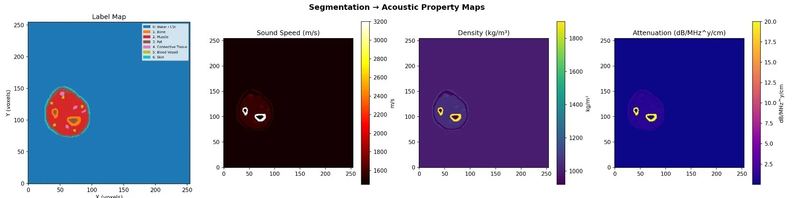
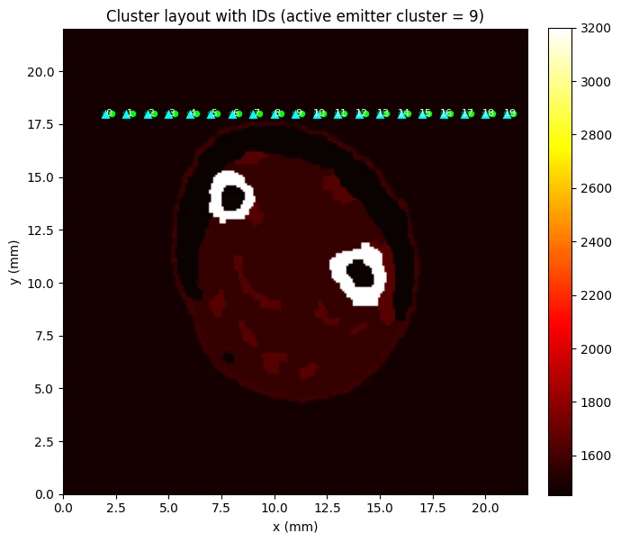
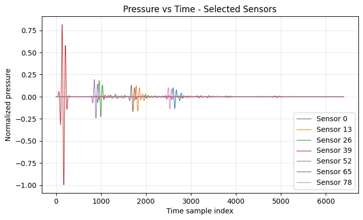
Select a stage to follow the workflow. Each figure comes from our project report; the ultrasound signals are simulated.
The approach
From anatomy to a signal.
We used forearm images from the USC Hand MRI dataset, segmented the tissue types, and assigned acoustic properties. These inputs were used in K-Wave simulations of ultrasound propagation.
We then extracted features—numerical summaries of the simulated signals—and used them to classify hand gestures. The study used four subjects, four poses, and two slices per pose, so the results represent an early proof of concept.
What mattered
A good score needs a good test.
The original evaluation produced near-perfect classification. But when the test data came from unseen subjects or source slices, accuracy dropped to around 32–39%, against a 25% chance baseline for four poses.
This difference was an important finding. Similar training and test data can make a model appear more capable than it is when faced with new anatomy.
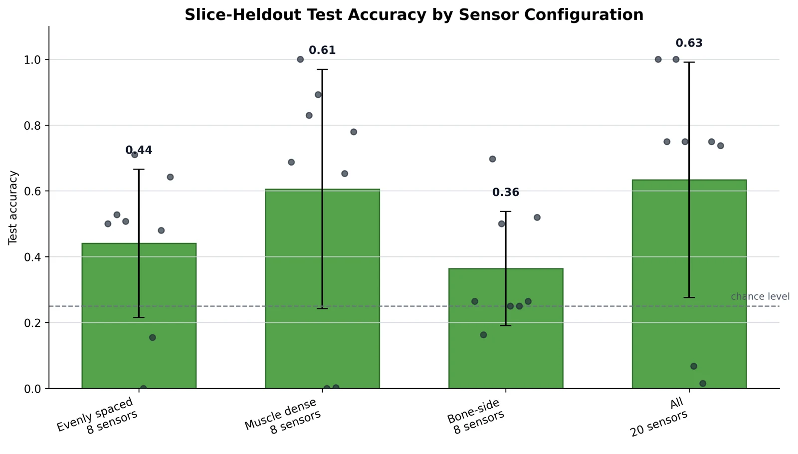
Later experiments with feature extraction and sensor placement improved performance. Eight sensors placed in muscle-dense regions reached around 61% average accuracy on slices kept out of training in the reported evaluation. The dataset remained small, with substantial variability between slices.
Sensor placement
Where you listen matters.
Sensor placement had a larger effect than tuning the tested classifier parameter. This links a machine-learning result back to the physical sensing arrangement.
Scope & team
An early proof of concept.
The work uses two-dimensional slices, static poses, literature-based acoustic properties, and synthetic temporal structure. It does not establish performance on real wearable ultrasound measurements.
Completed with Ifeoluwa Olusoji and Manuela Sanchez Gomez for P&O Nanoscience, Nanotechnology and Nanoengineering at KU Leuven.
Source: our 2025–2026 report, “From MRI to Synthetic Ultrasound for Hand Gesture Classification”. Anatomical inputs: USC Hand MRI dataset.